Compact Bone Diagram Endosteum / Illustration Of Endosteal Zone And Central Marrow Zone A The Endosteal Download Scientific Diagram - What is the difference between compact bone and cancellous bone?
Compact Bone Diagram Endosteum / Illustration Of Endosteal Zone And Central Marrow Zone A The Endosteal Download Scientific Diagram - What is the difference between compact bone and cancellous bone?. Each bone is an organ of the skeletal system. These bones tend to support weight and help movement. Learn vocabulary, terms and more with flashcards, games and other study tools. The endosteum is located on the internal surface of the bone, being the membranous layer that covers the medullary cavity, the bony trabeculae (spongy part of the bone), the haversian canals and internal walls of the compact long bones. It is a thin covering that surrounds it coats the inner compact bone and the trabeculae of the spongy bone.
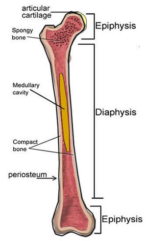
These cells add the compact bone to the bony callus to form a bone tissue that is similar to the original, normal bone. This endosteal surface is usually resorbed during long periods of malnutrition, resulting in less cortical thickness. To know the architecture of compact and spongy (cancellous) bone. Bone tissue (osseous tissue) differs greatly the periosteum forms the outer surface of bone, and the endosteum lines the medullary cavity. The outer shell of compact bone is called cortical bone or cortex.
These bones tend to support weight and help movement. The tissue that fills the spaces of the bone is called marrow 17. This endosteal surface is usually resorbed during long periods of malnutrition, resulting in less cortical thickness. Moreover, periosteum and endosteum cover the compact bone from outside and inner surface respectively. • a compact cortical shaft or diaphysis, (comprising a cylinder of compact bone, its cavity (medulla) being filled with spongy cancellous bone containing bone marrow). A similar layer, the endosteum lines the cavities. Cancellous bone is remodeled by endosteum. Compact bone is the cylindrical harder outer layer of the bone. In an adult, most red blood cells key terms. The densest and strongest bones in the body. Endosteum also has few connective tissues fibers and blood vessels. Definition and functions the endosteum is a structure in the middle of bone tissue and bone marrow. To know the architecture of compact and spongy (cancellous) bone.
Spongy bone, compact bone, articular cartilage, endosteum. It is found in bones such as the humerus and the. Learn vocabulary, terms and more with flashcards, games and other study tools. Membranes, including the endosteum and periosteum. A thin vascular membrane of connective tissue that lines the surface of the bone tissue that forms the medullary cavity of long.
6 3 Bone Structure Anatomy Physiology from open.oregonstate.education Compact bone diagram endosteum / solved question 20 secondary epiphysis d compact bone m chegg com : The outer shell of compact bone is called cortical bone or cortex. In other terms, they are the cortical bones; Flat bones are composed of two thin layers of compact bone that surround a layer of cancellous (spongy) bone. Cancellous bone is remodeled by endosteum. A thin vascular membrane of connective tissue that lines the surface of the bone tissue that forms the medullary cavity of long. It is a thin covering that surrounds it coats the inner compact bone and the trabeculae of the spongy bone. It covers the loose structures found inside the bone.
Flat bones, like those of the cranium, consist. What is the difference between compact bone and cancellous bone? In an adult, most red blood cells key terms. The two forms of marrow are red and. Compact bone is the cylindrical harder outer layer of the bone.
The endosteum (plural endostea) is a thin vascular membrane of connective tissue that lines the inner surface of the bony tissue that forms the medullary cavity of long bones.
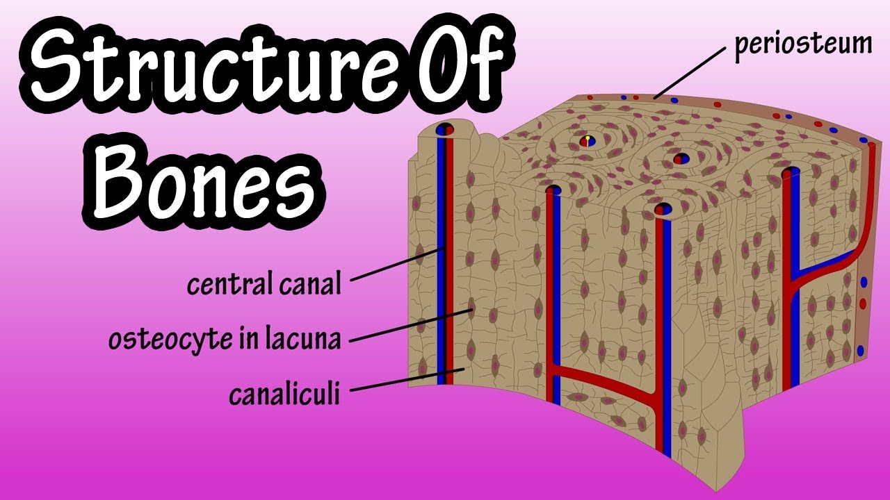
The osteoprogenitor cells of the preosteoblasts present in this connective tissue lining, differentiate into. Compact bone that forms the shafts of long bone consists of two structures. They are very difficult to distinguish from the surrounding connective tissue cells. Describe how bones are nourished and innervated. The endosteum is located on the internal surface of the bone, being the membranous layer that covers the medullary cavity, the bony trabeculae (spongy part of the bone), the haversian canals and internal walls of the compact long bones. Definition and functions the endosteum is a structure in the middle of bone tissue and bone marrow. Endosteum also has few connective tissues fibers and blood vessels. In this type of bone, the lamellae are organised into concentric circles, which surround a in both types of bone, the external surface is covered by a layer of connective tissue, known as the periosteum. The inset shows the lamellae of the compacta. Spongy bone, compact bone, articular cartilage, endosteum. The outer and inner regions contain layers of lamellar bone that run circumferentially around the entire bone. This endosteal surface is usually resorbed during long periods of malnutrition, resulting in less cortical thickness. The endosteum lines this cavity and endosteum contains bone forming cells 16.
Each bone is an organ of the skeletal system compact bone diagram. The outer shell of compact bone is called cortical bone or cortex.


0 Komentar